Diagnostic Services
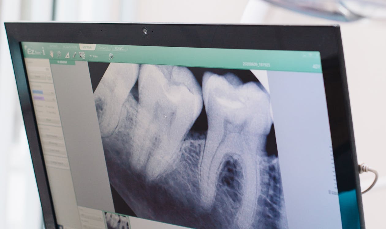
Diagnostic Images & Digital X-Rays
All diagnostic imaging at Wildwood Dental is captured digitally. Digital x-ray systems produce clear, immediate images that can be reviewed with you at your appointment and stored as part of your permanent dental record.
What digital diagnostic imaging means for you
Digital x-ray systems have replaced film-based technology at modern dental clinics. Rather than developing x-ray film in a darkroom, digital sensors capture images electronically and display them on screen within seconds. This changes the experience for patients in several practical ways.
Images are available immediately, so your dentist can review findings with you in real time, pointing to specific areas of concern and explaining what they show. There is no waiting, and nothing is left to memory or verbal description alone.
Digital images are stored as part of your electronic dental record and can be accessed at any future appointment, making it straightforward to compare current findings against prior images to track changes over time.
Radiation and digital x-rays
Digital x-ray systems use significantly less radiation than older film-based technology, typically 70–90% less. The sensors used in digital systems are more sensitive to radiation than film, so a much smaller dose is required to produce a diagnostic-quality image.
At Wildwood Dental, x-rays are taken based on clinical need, not on a fixed schedule. The frequency and type of imaging recommended depends on your individual history, risk level, and what is being assessed. We follow evidence-based guidelines and will always explain why a particular image is being recommended.
Types of diagnostic images we use
Diagnostic records at Wildwood Dental may include several types of images depending on what is being evaluated:
- Digital bitewing x-rays — The most common type, taken to detect cavities between teeth and assess bone levels in the back of the mouth.
- Periapical x-rays — Show the full length of one or more teeth, including the root and surrounding bone. Used to investigate specific concerns such as infection, root fracture, or bone changes.
- Panoramic x-rays — A single image that captures the entire mouth, covering all teeth, both jaws, and surrounding anatomy. Useful for new patient records and monitoring changes across the full arch.
- Clinical photographs — Standardized photos of the teeth, gums, and bite used to document baseline appearance and track changes in soft tissue or restorations.
- Intraoral camera images — Close-up images captured inside the mouth during examination, useful for documenting specific findings and showing patients what their dentist is seeing.
Accessing and transferring your records
Your diagnostic images belong to you. If you are being referred to a specialist, moving to a new provider, or seeking a second opinion, we can provide copies of your records. Digital records are straightforward to export and share with other dental or medical providers when needed.
To request a copy of your records or to have them sent to another provider, contact our clinic directly and we will assist you.
Ready to get started?
Call 306.374.7272 or request an appointment online.